Ultrasound carotid and vertebral arteries

The carotid arteries and vertebral arteries are the main arteries that supply blood to the brain. The common carotid arteries – one on each side of the neck - are arteries that originate in the aortic arch and reach the skull and brain through the cervix. Each common carotid artery is divided into external and internal carotid arteries which supply blood to the face and brain respectively. The internal carotid artery is considered more important than the carotid arteries, because its main function is to supply the brain with blood, and therefore with oxygen.
The condition that concerns us most in terms of carotid pathology is atherosclerotic or vascular or obstructive carotid disease, which usually affects people over 60 years of age and is more common in men than women, without excluding its occurrence in younger ages.
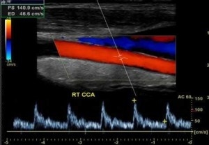
The colored Doppler ultrasound or Triplex of the carotid arteries and vertebral arteries is an easy, non-invasive, fast, painless and completely reliable imaging examination that enables us to check the condition of the carotid arteries in real time. With the Carotid Triplex we examine the anatomy and course of each vessel, the blood flow to the brain, the existence of atherosclerotic plaques and the extent, size, texture and composition of the atherosclerotic plaque. We can also visualize any stenosis and calculate with great accuracy the percentage of stenosis, either by measuring the percentage of free lumen, or by analyzing the spectrum and velocities of blood flow. Here we must say that a percentage of stenosis up to 40% is not considered hemodynamically important, it does not seriously disrupt the perfusion of the brain and is treated with medication, while as this percentage increases and especially when it exceeds 70-80% then the possibility of an invasive or surgical treatment of the stenosis either by carotid endarterectomy or by carotid angioplasty with stent placement.


