Image
Image

The arteries carry oxygenated blood from the heart to the rest of the body. The veins return the blood (poorer in oxygen) back to the heart.

There are three groups of veins in the lower limbs:

  • superficial veins (ie, the surface) that are closest to the skin,
  • the deep veins (that is, the deep veins) located within the muscle groups and
  • the perforated (or perforated) veins that connect the superficial veins to the deep veins.

Varicose veins are easily diagnosed by a specialist based on the clinical picture and examination with a portable Doppler. He is responsible for deciding whether a more detailed examination with a Triplex or venipuncture of the member's veins needs to be supplemented and what questions need to be answered with it. The most accurate studies are done by doctors specially trained in such examination.

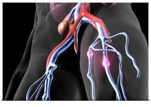
The carotid arteries and vertebral arteries are the main arteries that supply blood to the brain. The common carotid arteries – one  on each side of the neck - are arteries that originate in the aortic arch and reach the skull and brain through the cervix. Each common carotid artery is divided into external and internal carotid arteries which supply blood to the face and brain respectively. The internal carotid artery is considered more important than the carotid arteries, because its main function is to supply the brain with blood, and therefore with oxygen.

The condition that concerns us most in terms of carotid pathology is atherosclerotic or vascular or obstructive carotid disease, which usually affects people over 60 years of age and is more common in men than women, without excluding its occurrence in younger ages.

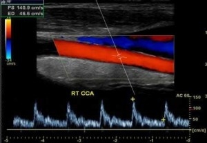
The colored Doppler ultrasound or Triplex of the carotid arteries and vertebral arteries is an easy, non-invasive, fast, painless and completely reliable imaging examination that enables us to check the condition of the carotid arteries in real time. With the Carotid Triplex we examine the anatomy and course of each vessel, the blood flow to the brain, the existence of atherosclerotic plaques and the extent, size, texture and composition of the atherosclerotic plaque. We can also visualize any stenosis and calculate with great accuracy the percentage of stenosis, either by measuring the percentage of free lumen, or by analyzing the spectrum and velocities of blood flow. Here we must say that a percentage of stenosis up to 40% is not considered hemodynamically important,  it does not seriously disrupt the perfusion of the brain and is treated with medication, while as this percentage increases and especially when it exceeds 70-80% then the possibility of an invasive or surgical treatment of the stenosis either by carotid endarterectomy or by carotid angioplasty with stent placement.

It is a non-invasive test used to assess the testicles and epididymis.

It is performed with an ultrasound machine, to which a special head has been fitted that allows the examination of these organs.

Most of today's machines also have Doppler technology, which measures blood flow to the testicles. The examination is performed by a radiologist or urologist who is trained in ultrasound of the urinary system.

There are many reasons why your doctor may recommend that you have the test:

  • In the investigation of a palpable mass (such as cancer) or in testicular pain.
  • In inflammation of the testicles or epididymis.
  • If testicular torsion is suspected (Doppler will show no blood flow).
  • In the diagnosis of presence of fluid in the scrotum (hydrocele), blood (hemorrhage) or pus.
  • In infertility (such varicose veins).

The procedure that will follow is:

  • You will lay down on the examine bed, after removing your clothes from the middle and bottom, in a supine position.
  • A clear thick gel will be applied to the examination site. You should be aware that any residue left, does not stain your clothes.
  • The doctor will then apply the ultrasound head on the gel and begin to scan the area. You might be asked to take a deep breath and hold it to push it down, increasing the pressure on the abdomen.
  • This is done to evaluate the varicose vein and the regression of venous blood to the testicles.
  • After the test, the doctor will help to wipe excess gel.


Ultrasound soft tissue examination is a highly reliable method and involves muscles, tendons and subcutaneous tissue. Second- and third-degree traumatic muscle ruptures with concomitant hematoma are easily detected by ultrasound, as are tendon calcifications in chronic tendonitis, while small elongated and transverse ruptures may escape.

Baker cysts in the iliac cavity and ganglion cysts in the wrist are relatively easy to detect with ultrasound, but they often have difficulty in detecting injuries to the joints. The screening of tumors in the subcutaneous tissue, most commonly lipoma and sebaceous cysts, is done with great precision by ultrasound as well as the appearance of swelling in the subcutaneous tissue (lymphedema) or inflammation. The study of soft tissues with ultrasound, including also the nerves, resulting to achieve local anesthesia.

Contracted with EOPYY

Image

All major credit cards accepted

Credit or Debit cards
Image